The Outlaw Pad Laminitis Treatment
Horseshoe Related Articles
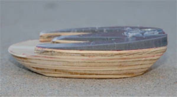
A good idea can come from any where.. these articles were published 6 months after the release of the OUTLAW SHOE.
Goals of Treatment for Chronic Laminitis
Quote: “Trimming and shoeing has always been the “mainstay” of treating chronic laminitis, and it is directed at reducing/removing the adverse forces on the compromised lamellae. In considering hoof care in horses with chronic laminitis, there are three goals for therapy: to stabilize the distal phalanx within the hoof capsule, to control pain, and to encourage new hoof growth to assume the most normal relationship to the distal phalanx possible. Realignment of the third phalanx to create a better relationship of the solar surface of the distal phalanx with the ground is used as the basis for treating chronic laminitis.
The same end can be achieved by cutting the thinner piece in the shape of a “W” and then attaching it to the thicker section of plywood as described above (Fig. 4).”
Stephen E. O’Grady, BVSc, MRCVS; Micheal L. Steward, DVM; and Andrew H. Parks, VetMB, MRCVS, Diplomate ACVS
The Blacksmith likes this article!
The Horse newsletter sent by: Tracy Gantz
November 17 2010, Article # 17254.
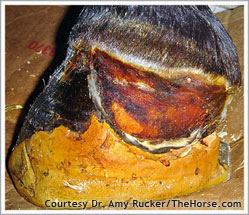
It shows the severity of an untreated foundered foot. Why not use an OPEN TOE HEART BAR SHOE from the onset of any laminitic or founder case?
When laminitis develops to the point that the hoof wall is restricting blood supply to the laminae and causing further inflammation, a hoof wall resection might be needed. Amy Rucker, DVM, of Midwest Equine in Columbia, Mo., spoke about when to do resections and methods that give veterinarians the best chance for success during the Sept. 17-18 Laminitis West Conference in Monterey, Calif.
In some laminitis cases, the distal (lower) rim of the coffin bone becomes septic (infected) due to compromised blood flow and dying tissues. Rucker explained that fluid resulting from this can travel up the lamellar interface, breaking out at the coronary band. If initial treatment, usually a poultice, doesn’t stop the process, the coronary band will swell and prolapse over the hoof wall. The wall then cuts into the swollen tissue, causing further inflammation and restricting blood supply to that tissue.
Before Treatment for Laminitis
After Treatment for Laminitis
This hoof needed a partial resection due to coronary prolapse on one side (this photo was taken three weeks after resection).
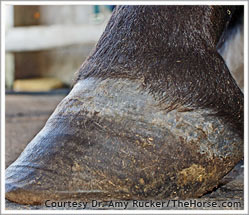
About nine months later, the hoof has grown out completely and the horse is sound.
Performing a resection–literally taking off part or all of the hoof wall–seems drastic, but it can relieve the pressure on the laminae and thereby allow the critical blood circulation to be restored. With meticulous aftercare, the foot’s underlying tissues can then repair.
“You don’t want to let these cases get away from you,” Rucker said. At the first sign of coronary prolapse, she re-evaluates her treatment and makes shoeing adjustments to change the loading pattern within the laminitic foot (a deep digital flexor tenotomy might also be considered; for more information, see Webinar: Laminitis Diagnosis and Treatment). She also medicates the coronary band twice daily with a poultice. Most cases respond in two to three days (drainage and swelling quickly resolve), but non-responsive cases require resections.
Rucker noted the following aspects of her technique for performing hoof wall resections:
- She removes the desired portion of the wall by cutting through it in a gentle arch/smiley face pattern, extending that past the coronary band prolapse and down below the affected tissue. She slowly pulls the wall off, easing apart the coronary papillae and lamellae.
- After removing the portion of the wall, she checks the remaining wall to make certain no sharp edges remain.
- The coronary and lamellar tissues are evaluated for degree of compromise. Mild cases will have some edema or swelling, but when the lamellae are gently massaged they begin to bleed (indicating the circulation has returned). Cases with moderate damage have unhealthy-looking coronary and lamellar tissue and may require more aggressive treatments such as the application of platelet-rich plasma. Cases with long-term or severe damage may have areas of devitalized tissue at the coronary band or lamellae and the affected portion of the coffin bone may be evident. Any exposed bone can be cut away and bandaged.
- Rucker cuts a piece of felt (cast, automotive upholstery, or carpet felt) to the exact size and shape of the resected wall. She soaks it in Betadine and bandages it tightly over the defect to minimize swelling. The bandage is changed daily, and new areas of epithelialization (growth of cells over the resected area) should be evident within a week. The defect should begin to cornify (harden) within seven to 10 days and will eventually grow out as new hoof wall grows down from the coronary band. If treatment is successful, the white line of the new wall will be visually indistinguishable from that of a normal horse.
- Aftercare, especially keeping the foot tightly bandaged, is critical, Rucker said, as is aggressively treating cases at the first sign of swelling or drainage from the coronary band. Waiting too long for resection results in irreversible damage to the tissue. The wall may grow back, but only have a weak attachment to the underlying lamellae and coffin bone. In addition, the new wall itself may be disorganized and weak, lacking tubular horn if its growth center at the coronary band is damaged.
- “As with all cases of laminitis, the key to success is immediate treatment,” she said. “Feet with minimal bone damage that can establish a strong bone-lamellar-wall attachment recover and return to soundness, but owners must be patient as it takes a year for the feet to regrow. Owners must also address the underlying cause of laminitis. In our area of the country, that is often obesity combined with insulin resistance.”